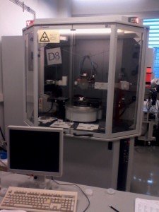
XRD: BRUKER D8 ADVANCE HIGH RESOLUTION DIFFRACTOMETER
X-ray scattering is routinely used to determine the crystal structure, orientation, lattice parameters and crystal quality in crystalline materials (X-ray diffraction) and thickness, density or roughness of thin films and multilayers (X-ray reflectivity).
The equipment configuration is optimized for high-resolution X-ray diffraction (HR-XRD) and reflectivity (XRR) studies in nanostructured thin films and superlattices. For this purpose it includes incident and diffracted-beam monochromators, collimators, attenuators and eulerian sample holder. It also allows the local mapping of flat samples through motorized lateral displacements.
XRD at LMA uses a D8 Advance diffractometer provided by Bruker Española S.A. This instrument has several modes, including:
- High resolution diffraction in epitaxial films and multilayers
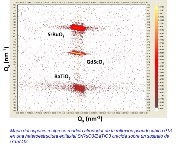
- Reciprocal space maps
- Wafer mapping
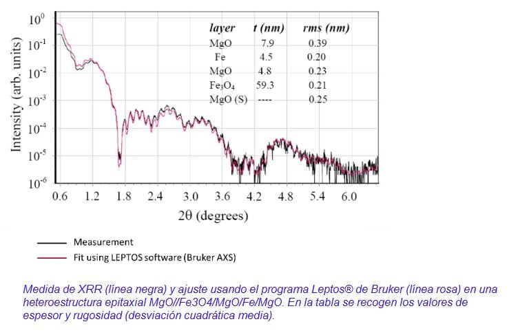
- X-ray reflectivity
- Structural characterization of single-crystal or strongly textured thin films
- Grazing incidence diffraction (GID)
The XRD equipment is mainly used by researchers growing epitaxial thin films in the following applications and research lines:
- Epitaxially strained thin films.
- Magnetic thin films and nanostructres for applications in Spintronics.
- Multiferroic films and multilayers.
- Thin films showing thermoelectric effects (such as spin Seebeck).
What can be done with it?
XRD and XRR provide the following information in thin films:
- Chemical composition.
- Crystal structure.
- Lattice parameters.
- Substrate and film orientation.
- Miscut angle in single-crystal substrates.
- Crystalline quality and texture.
- Roughness.
- Density.
- Thickness.
- Defects, dislocations.
- Stress.
Sample requirements:
Although this facility is very versatile and the analysis of powder samples is feasible, the current configuration is optimized for the study of thin films.
Technical specifications:
- X-ray generator with copper anode.
- (022) Ge monochromator (Cu Ka1 line).
- Parallel-beam optics (Göbel mirror).
- Eulerian cradle and XYZ translation stage.
- Zeta and Xi tilt stage for grazing incidence X-ray diffraction.
- Suction device for securing flat samples.
- Soller slits.
- X-ray beam intensity attenuator.
- Automatic alignment control by microscope and laser beam.
- Scintillation counter.
- Analysis software and databases.