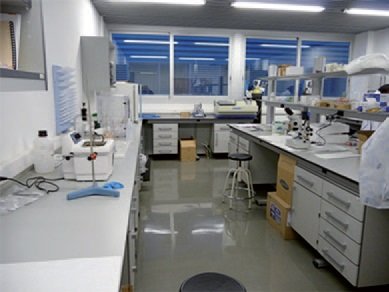
TEM SAMPLE PREPARATION SERVICE – LMA
Transmission Electron Microscopy (TEM) is based on the use of transmitted electrons to form images of the materials. However, the electron is a strongly interacting charged particle and TEM samples are required to be extremely thin (tens of nm) to be electron transparent. Some materials are inherently electron transparent (nanoparticles, nanotubes…), but most of them (bulk materials, thin films, devices…) have much larger dimensions and it is frequently required a TEM sample preparation procedure to make them thin. The LMA has set the complete Sample Preparation Laboratory equipped with the necessary instruments to perform this task.
Among the many procedures to produce electron transparent specimens, the most important and frequently used is based on mechanical thinning of the materials in a highly controlled way. This produces a flat specimen of a few microns thickness with smooth surfaces that afterwards follows a low-angle, low-energy ion milling of the surfaces to achieve extremely thin areas ready for TEM observation.
Ultramicrotomy is another technique to produce TEM samples of soft materials. The procedure consists in cutting some slices of the sample using the diamond knife of an ultramicrotome. These slices are around 50 nm of thickness of polymers and biological samples. Depending on the specific features of the materials, samples can be cut between -180ºC and room temperature. In the case of biological samples, a previous treatment is necessary in which the samples (tissues, cells, bacteria…) need to be fixed (using glutaraldehyde and osmium tetroxide), dehydrated and embedded in epoxy resin before cutting with the ultramicrotome.
In the LMA it is possible to vitrify samples and analyze them in cryogenic conditions. In this case, aqueous solutions like macromolecules, proteins, viruses… can be vitrified using liquid ethane which, after this, can be observed using cryo-TEM technique.
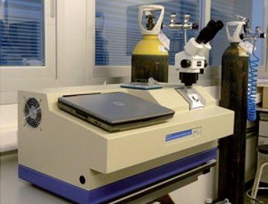
Instruments available for TEM sample preparation:
- Ion Mill Fischione Model 1010.
- Plasma Cleaner Fischione Model 1020.
- Low/high-speed polishers.
- Tripods and grinders.
- Dimpler.
- Diamond wire saw.
- Stereographic microscope.
- Metallographic microscope.
- Inverted microscope.
- Ultramicrotome Leica EM UC7.
- Cryo-ultramicrotome Leica EM FC7.
- FEI Vitrobot (vitrification).
Materials that can be prepared for TEM observation:
- Nanoparticles, nanotubes,…
- Inorganic materials (oxides, metals, ceramics) in cross-section and plane view).
- Polymers.
- Organic molecules (proteins, DNA, gels, virus, …).
- Cells and biological tissues.