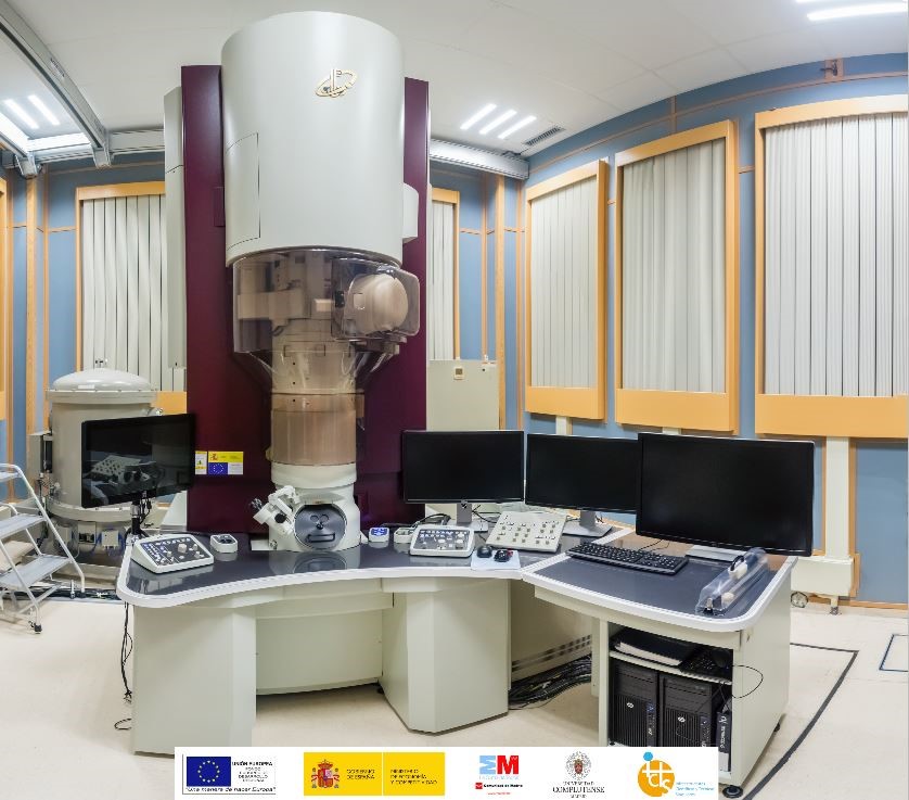
JEM GRAND ARM 300cF
The JEM GRAND ARM 300cF in the Centro Nacional de Microscopía Electrónica, CNME, is unique in Spain. The configuration chosen was aimed towards an electron microscope to be operated in Conventional Transmission Electron Microscopy, CTEM. It is then specially indicated in everything related to crystal-chemistry analysis of materials. However, the microscope also has attached spectrometers for XEDS and EELS, oriented to complete the structural information.
- The ETA CORRECTOR is mounted in the objective lens of the microscope and is able to correct aberrations up to the fifth order. The operation of the corrector, that is measuring aberration and applying the subsequent improved values to the lenses in the corrector, is quite user friendly through the use of the software COSMO.
- The cathode is Cold Field Emission Gun which delivers a high intensity beam with a very low energy spread.
- Even though the microscope is specially design form CTEM, it also has STEM detectors both for dark and bright field imaging. The possibility of recording ABF images is also there as long as there exits a beam stopper.
- This microscope has a newly design and implemented vacuum system (based on the use of turbo molecular pumps) which is specially oriented to deliver a highly clean atmosphere inside the column.
- It has available alignments at 60, 80, 120, 200 and 300 kV. This opens up the possibility to play around with the acceleration voltage as a parameter to improve the stability of the samples. Additionally, the electron dose can be also adjusted as the microscope has up to 8 condenser apertures with different sizes.
- A very fast CMOS OneView digital camera which offer a wide field of view (4Kx4K). The camera allows to correct for the drift in-situ and has the remarkable ability of recording images with very short exposure time, well below 0.1 s.
- The CENTURIO XEDS detector is a quite efficient one which has been optimized to offer a fast and highly sensible collection through the use of a wide solid angle collection and high active area.
- The ENFINIUM spectrometer for EELS analysis is very user friendly at moderately high resolution.
- The microscope is used for very high resolution (up to 0.045 nm) work. Thus, the analysis of complex crystal structures is the main target to tackle with this microscope due to the point to point resolution that can be obtained as a consequence of the corrector placed at the objective lens. It is possible to obtain images of atomic columns in advanced functional materials of very diverse composition (semiconductors, superconductors, magnetoresistant, dielectrics, multiferroics…), even of light atoms, allowing the identification of distortions, atomic displacements, order-disorder phenomena, both in cationic and anionic sublattices, superstructural ordering, domain structures and, in general, everything related to defects in solids, which are intimately connected to the physical and chemical properties. The applicability of the microscope must not be reduced to these aspects within solid state sciences but it is also becoming important in areas related to macromolecular sciences. Catalysis, organic chemistry, biology, etc. are well within the areas that are now of interest for this microscope.
- Closely connected with what it has been stated in the previous point, nowadays is very relevant the use of electron microscopes to study compounds that are beam sensitive. In this respect the microscope is optimized to work at 60 and 80 kV. Good examples are the studies that these days are been done in carbon nano-structures with sub-Ångstrom resolution. Note that these type of studies are not reduced to carbon nano-structures but also in compounds like hybrid organic-inorganic, low dimensional materials, metal organic frameworks, proteins, and so on (Figures 1 to 3).
- The microscope is equipped with the STEM unit along with dark and bright field detectors. Then, the information out of the ultra-high CTEM studies can be completed and/or combined with the information from the STEM studies. These together with the XEDS and EELS spectrometers allows a very complete analysis of the samples covering all aspects of interest: atomic structure, composition and bonding altogether.
- Cold field emission gun (cFEG).
- Vacuum system comprising 6 ionic and one turbomolecular pumps.
- Aberration corrector ETA (Expanding Trajectory Aberration) located at the objective lens: aberration compensation via two dodecapoles. It contains certain elements capable of reducing chromatic aberrations.
- JEOL COSMO Software to measure aberrations, and to adjust the optical system.
- Column designed to minimize image distortions caused by temperature and magnetic fields fluctuations.
- High tension stability: 0.4 ppm (P-P)
- Objective lens electric current stability: 0.5 ppm (P-P)
- Acceleration voltages: 300, 200, 120, 80 and 60 kV.
- Objective lens (TEM): ultra-high-resolution configuration (FHP)
- Focal length 0.7 mm, Cs 0.7 mm, Cc 1.3 mm, minimum focus step 0.3 nm.
- Objective lens (STEM) Cs -0.1 a 0.6 mm, Cc 1.4 mm.
- Scanning system in transmission mode (STEM) and DF/BF/LADF detectors.
- Magnification range (TEM): 2,000x – 2,000,000x.
- Magnification range (STEM) 20,000x – 150,000,000x
- Camera length range: 80 – 1500 mm.
- Specimen tilt range: ±25 degrees.
- Spatial resolution in TEM: 0.05 nm, point to point at 300, 200 y 120 kV; 0.1 nm at 60 y 80 kV
- Spatial resolution in STEM: 0.1 nm at 300 kV; 0.2 nm at 80 kV
- Gatan Quantum EELS Enfinium with an energy resolution of 0.55 eV.
- Gatan OneView camera: from 4096×4096 pixels at 25 fps to 512×512 pixels at 300 fps, for fast image acquisition and in-situ experiments. Image formats 1:1 (4k, 2k, 1k) 16:9 (UHD, HD). Video formats 1:1 (4k, 2k, 1k) 16:9 (UHD, HD)
- SDD CENTURIO X-EDS detector with an active area of 100 mm2, and a solid angle of 0,8 sr, with an ultra-high resolution polar piece of the objective lens.
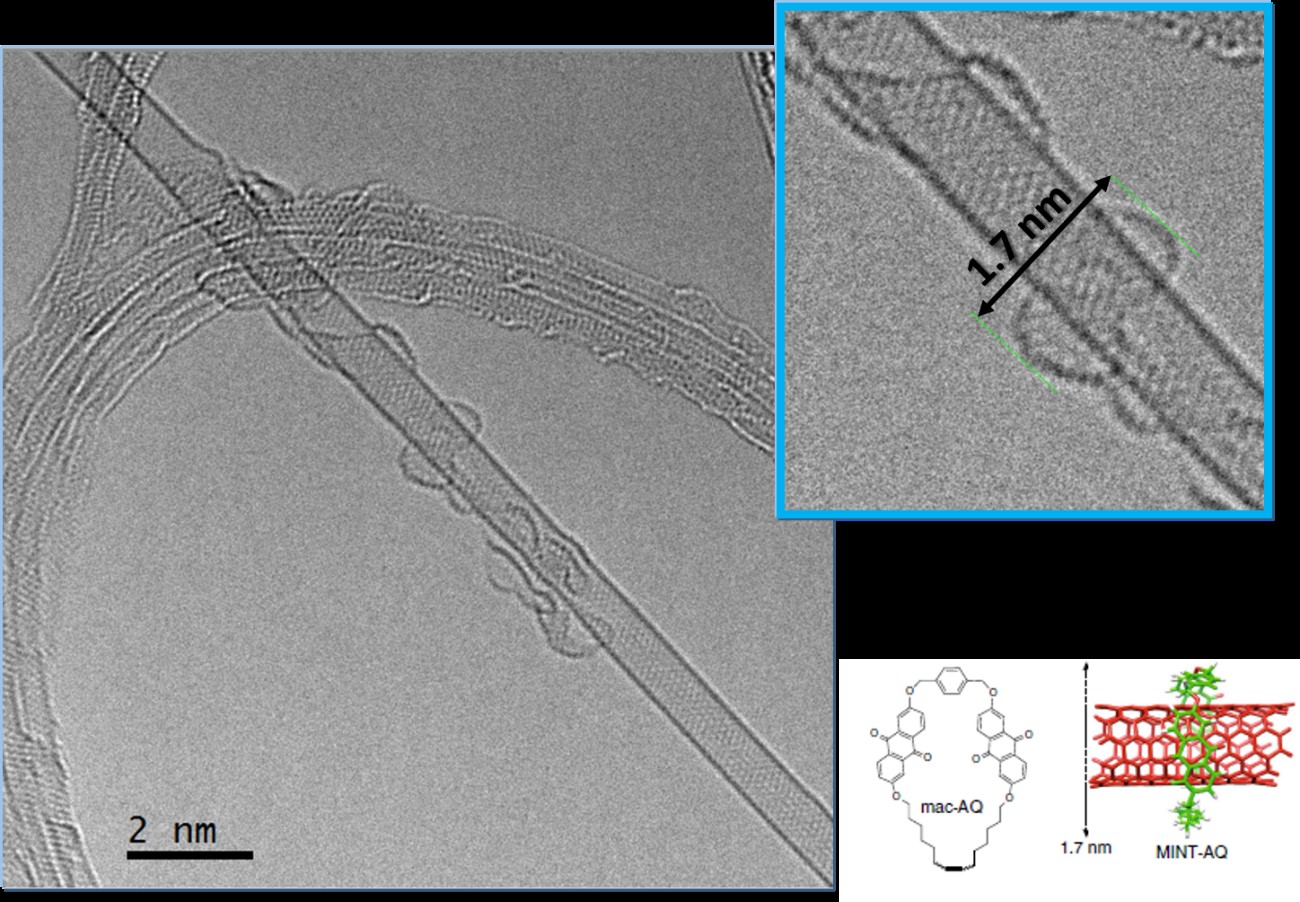
Figure 1. High resolution CTEM image recorded in the GRAND ARM cFEG. The microscope was operated at 60 kV. In the image we observe a MINT (Mechanically Interlocked Nanotube). The single wall nanotube is decorated on the surface with a macrocycle molecule. Figure from Nature Communications 9(2671), 1-7 (2018).
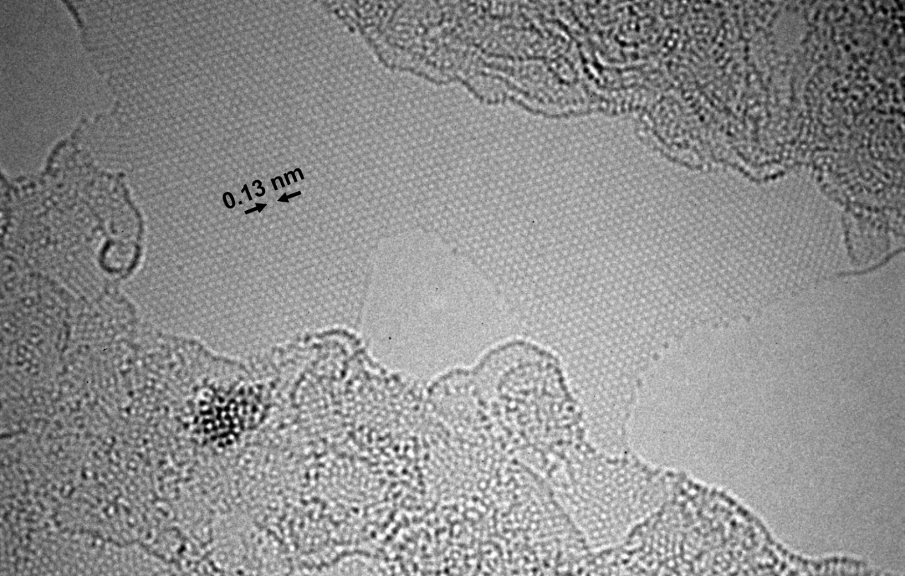
Figure 2. High resolution image recorded in the GRAND ARM300 cFEG operated at 60 kV. In the central area of the image a mono-layer of graphene is observed.
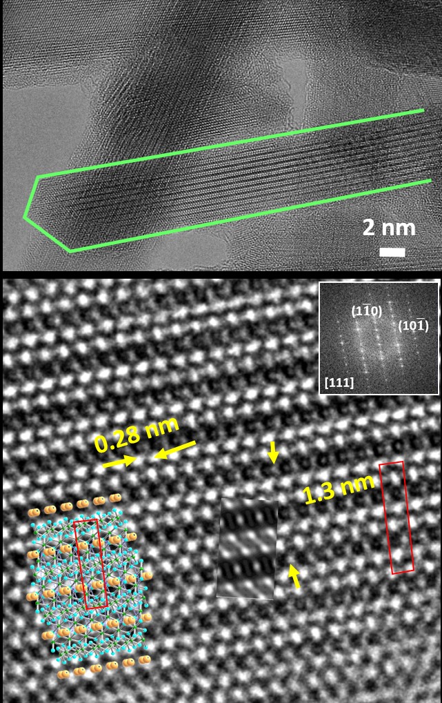
Figure 3. CTEM high resolution image recorded in the GRAND ARM 300 cFEG operated at 80 kV. The top panel shows an acicular related hollandite nanoparticle of composition K0.15MnO2+d. The bottom panel shows a higher magnification detail where the simulated image is inset, together with the structural model along the corresponding direction. Chem. Comm. 56, 4812 (2020).
E. Burzurí et al, “Magnetic, Mechanically Interlocked Porphyrin–Carbon Nanotubes for Quantum Computation and Spintronics”. Journal of the American Chemical Society, 143(50), 21286 (2021).
E. Pérez et al, “Covalent modification of franckeite with maleimides: connecting molecules and van der Waals heterostructures”, Nanoscale Horizons, 6, 551, (2021).
J. Puigmarti-Luis et al, “Biomimetic Synthesis of Sub-20 nm Covalent Organic Frameworks in Water”, Journal of the American Chemical Society 142, 3540 (2020) .
A. Castellanos-Gómez, “Mechanical and liquid phase exfoliation of cylindrite: a natural van der Waals superlattice with intrinsic magnetic interactions”, 2D Materials, 6(3),035023-1, (2019).
S. Casado et al, “Reversible dispersion and release of carbon nanotubes via cooperative clamping interactions with hydrogen-bonded nanorings”, Chemical Science 9, 4176 (2018).
H. Sawada et al, “Positive and negative regulation of carbon nanotube catalysts through encapsulation within macrocycles”, Nature Communications 9(2671), 1 (2018).
D. M. Guldi et al, “Interfacing porphyrins and carbon nanotubes through mechanical links”, Chemical Science 9, 6779-6784 (2018).
M. J. Mancheño et al, “Thiol grafted imine-based covalent organic frameworks for water remediation through selective removal of Hg (II)”, Journal of Materials Chemistry A 5, 17973 (2017).
E. M. Pérez et al “Band-Gap Opening in Metallic Single-Walled Carbon Nanotubes by Encapsulation of an Organic Salt”, Angewandte Chemie Int. Ed. 56, 12240 (2017).
F. Zamora et al. “Ionic Conductivity and Potential Application for Fuel Cell of a Modified Imine-Based Covalent Organic Framework”, Journal of the American Chemical Society 139, 10079, (2017).